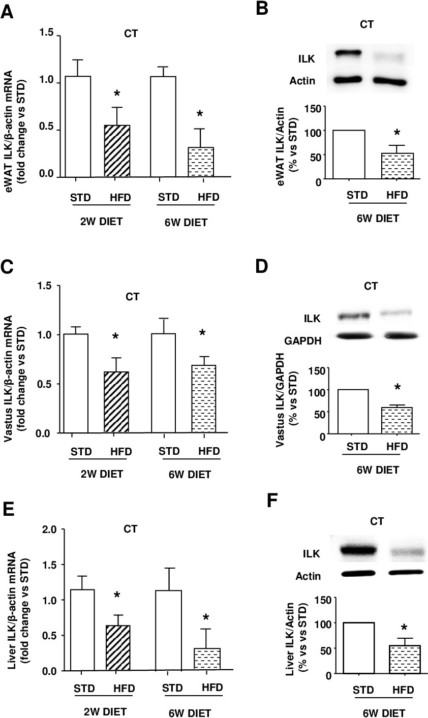
Fig. 3. ILK expression in adipose, skeletal muscle and hepatic tissues from CT during short-term STD or HFD challenges. Fresh isolated epididymal white adipose tissue (eWAT), vastus and liver explants were extracted from CT fed with either STD or HFD for 2 or 6 weeks (w). Animals were maintained in fasting conditions before to extract the tissues and proceed to the determinations. ILK mRNA expression levels fold changes, analyzed by RT-qPCR and normalized to β-actin as endogenous control, in eWAT (A), vastus (C) and liver (E) from CT fed with either STD or HFD for 2 or 6 weeks (w). Representative immunoblots and densitometric analysis of ILK protein levels normalized to Actin or GAPDH as endogenous control from eWAT (B), vastus (C) and liver (E) of CT fed with either STD or HFD for 6w. ILK expression transgenically downregulated in basal cKD-ILK tissues has been previously shown (Hatem-Vaquero M et al. J Endocrinol. 2017 Aug;234(2):115-128). N=12 per group. All data are represented as means ± SEMs. *= P<0.05 vs STD at the same time point.